
In our first cavity series we showed you what an actual cavity looks like on an X-Ray and in a real life photo. Both of the cases were of the back teeth. Now we bet you are curious – what do cavities look like on front teeth?
Actual Photos
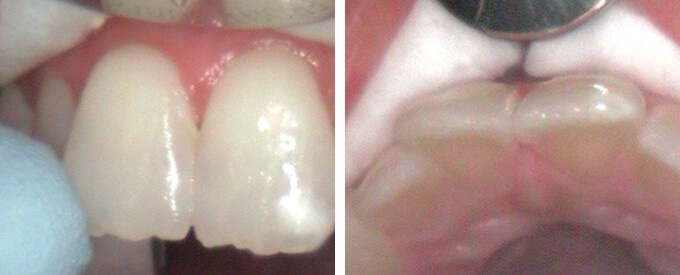
These two pictures show the before photos of an individual’s upper two front teeth. At first glance they may look completely normal to you! It is only when the dentist looks at the X-Ray and then at the back of the tooth (tongue side) that we can see the cavity between the teeth.
X-Ray on Front Teeth
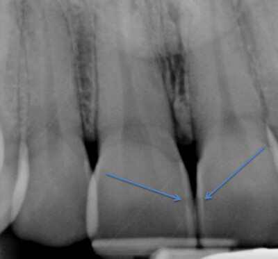
In between the blue arrows you can see a small dark shadow. If you remember from the first blog, that indicates a cavity is present. The enamel of the tooth has been destroyed and the bacteria has gained access to the inside of the tooth.
The “In-Between” Photo
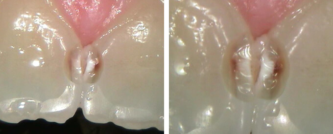
The cavity was removed from the back side of the teeth to maximize the esthetics/front view of the teeth. The dark brown area clearly shows you the cavity destroying the tooth and the white chalky area is the damaged enamel.
Filling Replacement & Final Photo
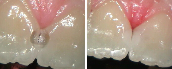
The first photo shows the teeth after removing all of the damaged tooth and the area is now ready for the filling. The second photo shows the teeth completely restored using a tooth-colored composite material.
Now you may see why it is always important to take an X-Ray at your every six-month cleaning appointments!

