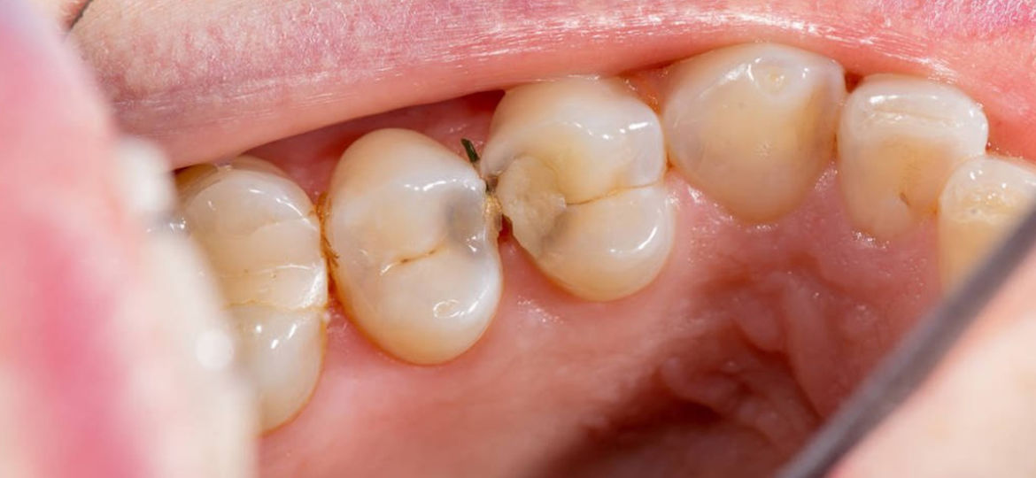
Many of you ask us, what does a cavity actually look like in your tooth? In a series on cavities, we will show you what they look like and how we fix them. In this particular case, we will show you the cavity on an X-Ray, an actual photo of one being fixed and then lastly the filling in the tooth.
X-Ray Photo
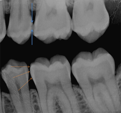
The blue arrow shows the cavity. It is a dark shadow between the arrows. This shadow represents a “hole” or “tunnel” through the protective enamel of the tooth where bacteria now has access to the soft inside portion of the tooth. The orange arrow shows an area of normal/healthy enamel. There is a solid white consistency with now shadows. Do you see the difference?
Picture of Cavity
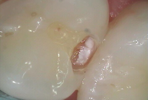
This picture was taken after cutting a small hole in the tooth to access and repair the cavity. The chalky white area in the photo is the damaged enamel of the tooth and the chalky looking area corresponds to the dark shadow look on the X-Ray.
One Year X-Ray
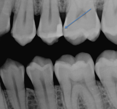
The blue arrow shows the filling after one year and as you can see it shows up bright white on the X-Ray. This means the cavity is still in good order!
Example Two
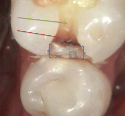
We also have another example for you where the cavity is bigger and more destructive than the first example. The blue brackets show the enamel in between the teeth that has been damaged. You can see the whitish/chalky appearance to this enamel and also the crack in the middle of the tooth.
The red arrow indicates the discolored brown inside of the tooth. This brown area is what a cavity looks like once it gets through the enamel layer and starts destroying the inside of the tooth. The green arrow is an example of what the inside of a normal healthy tooth should look like – it should be slightly yellowish in color.
Before and After
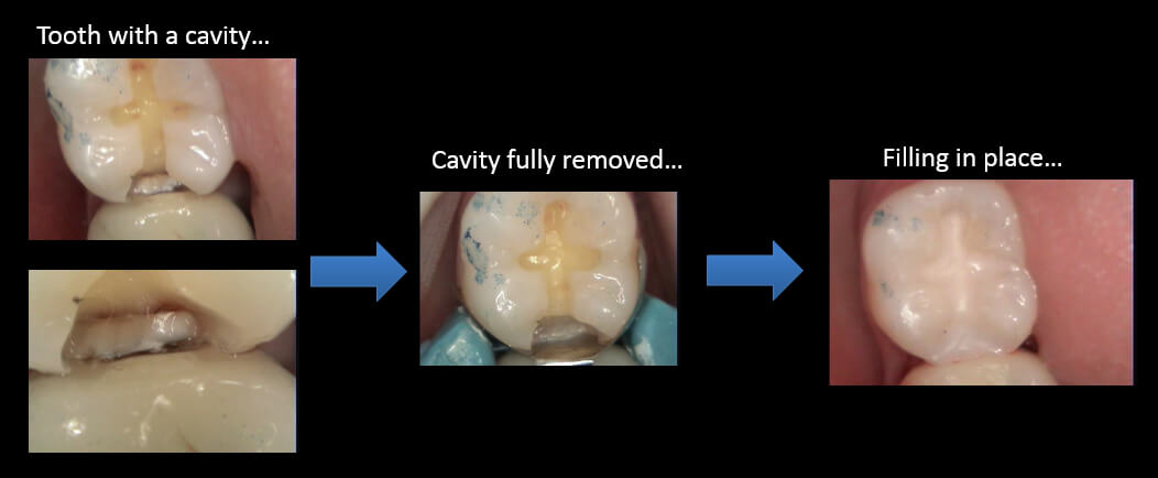
And lastly photos from start to finish on a cavity! Keep an eye out for a few more stories about cavities in this series!

