Do you dread hearing the words “root canal”, but you’re not sure why? For some reason the term can strike a moment of fear even in the bravest of individuals. We’re here to tell you… Fear Not!!
This particular case will give you more information about root canals, why they are important and how they can actually help keep you pain-free if you ever have a toothache.
X-Ray
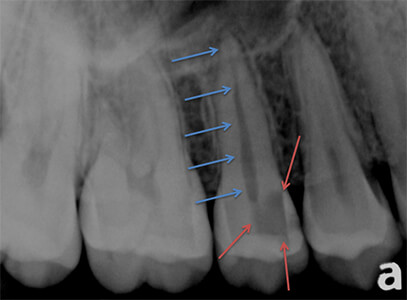
The X-Ray shows a tooth with a very large cavity. It has actually reached the nerve of the tooth:

- Blue arrows – point to where the Nerve Canal of the tooth is
- Red arrows – show where the cavity is.
You can see the the cavity is touching the nerve. This causes the nerve to die and fill with bacteria and infection, which causes the painful toothache.
So, the question is how do we fix this?
Root Canal Photos

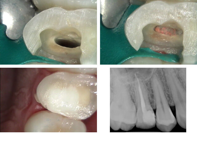
The first picture shows what the tooth looks like after removing the entire cavity and then removing all of the infected nerve tissue.
The second picture shows the root canal filling material in place (orange colored stuff in the middle).
The third picture shows the core buildup in place restoring the missing tooth structure where the cavity used to be.
Finally, the last picture is the x-ray of the completed root canal and core buildup.
Final Step: Crowning the Root Canalled Tooth

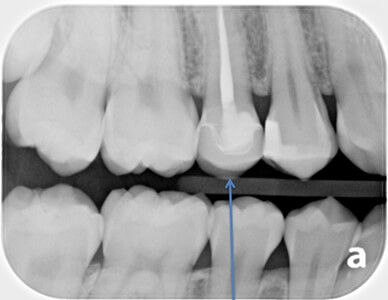
The final X-Ray shows a crown on the root canalled tooth. This particular tooth had a very large cavity and is considerably weakened because of all the missing tooth structure. The crown is very important to prevent this tooth from fracturing. The root canal procedure allowed the dentist to SAVE this persons tooth and to prevent a painful toothache. Without a root canal, this tooth would have needed to be extracted.
Contrary to popular belief – root canals are NOT painful… they are comparable to having any other minor dental procedure done (a filling, etc). If you currently have a toothache and are nervous to find out if you need a root canal – don’t be! Give our office a call today and we are happy to help.

