There are numerous ways that 3D imaging is used in a modern dental practice. This page will share with you some of the amazing things this technology can do.
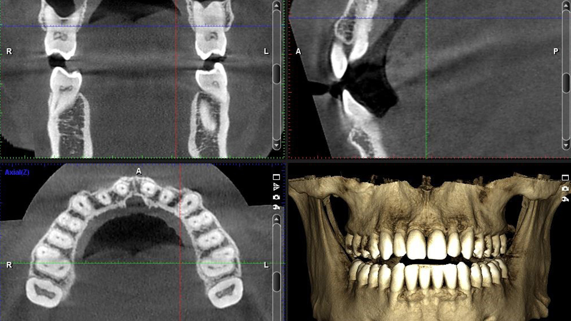
The Benefits of 3D Imaging
3D imaging allows your doctor to see “inside out”. Imagine if you could see the inside of an apple, but without ever having to cut the apple in half? This is what CT allows us to do.
Implant Planning
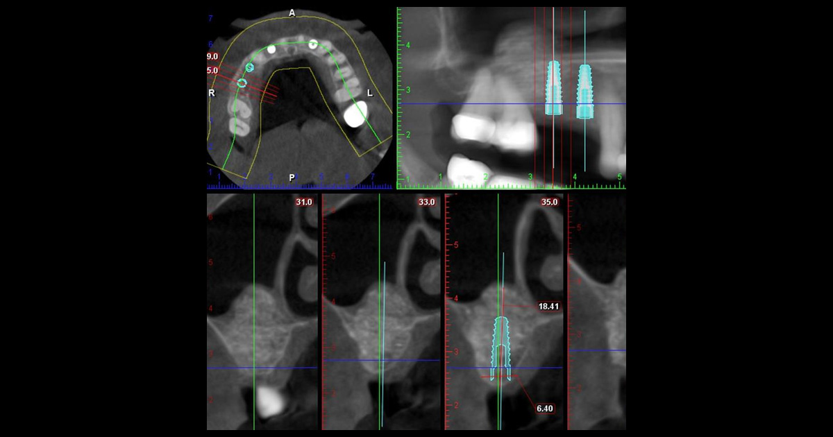
Before any treatment – CT used to diagnose if the patient is a candidate for implants:
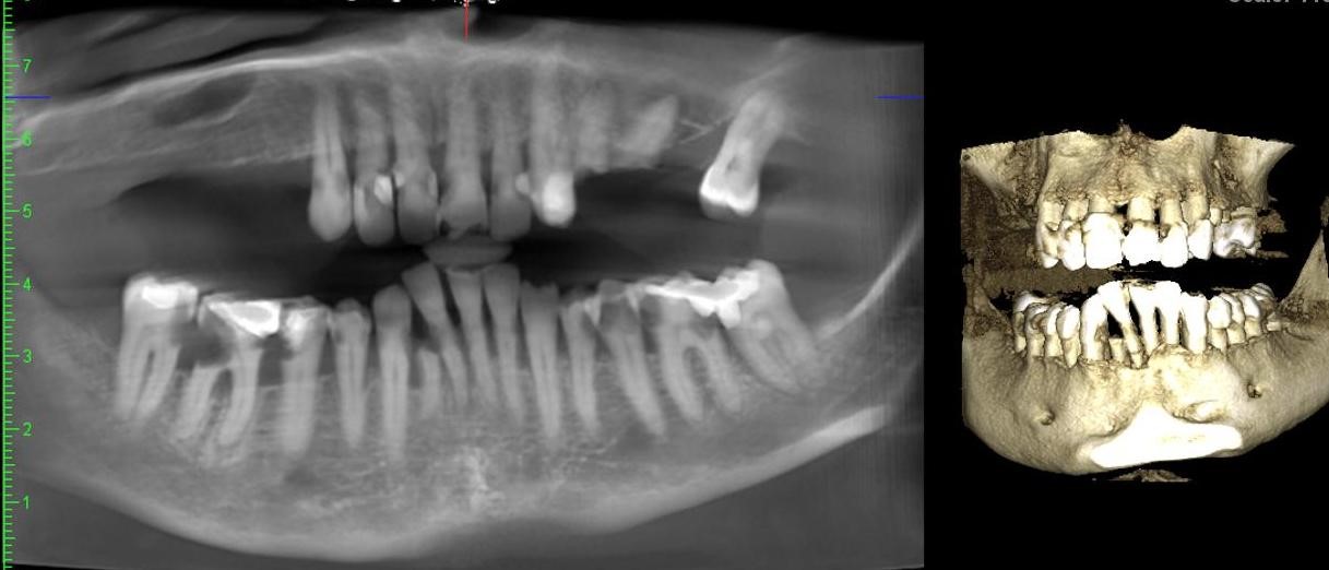
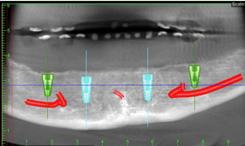
After extracting teeth and making denture – CT used to plan four implants on the lower jaw:
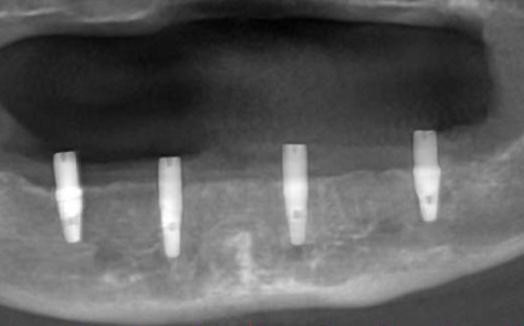
After implants placed – CT used to verify precise placement of implants within the bone:
Root Canal Planning
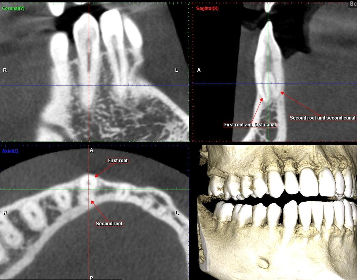
Sometimes teeth have extra canals – and often even entire extra ROOTS – that cannot be seen using normal 2D imaging. In this example, the 3D image shows how this lower canine has an extra root and an extra canal. With this technology, the dentist knows about this abnormal anatomy BEFORE the procedure. This makes root canal treatment more predictable and more successful.
Diagnosis of Pathology
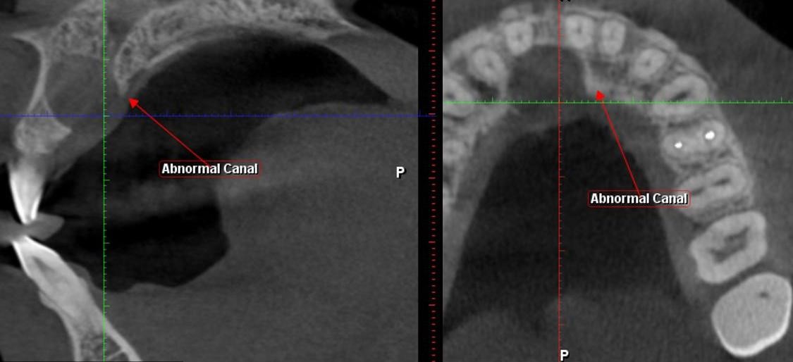
3D imaging is very useful in diagnosing maxillofacial pathology. Sometimes your dentist may see something on a 2D image and not know what it is. 3D imaging allows us to get a better view. Below we’ll use a certain type of pathology called an “Incisive Canal Cyst” as an example. These examples are all from our office.
After implants placed – CT used to verify precise placement of implants within the bone:
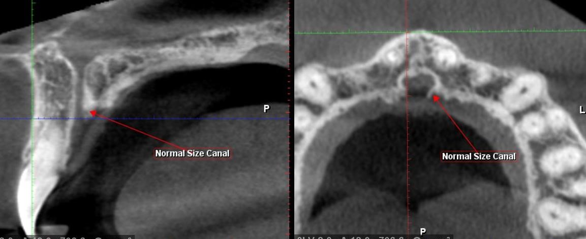
Example #1 of what a cyst looks like:
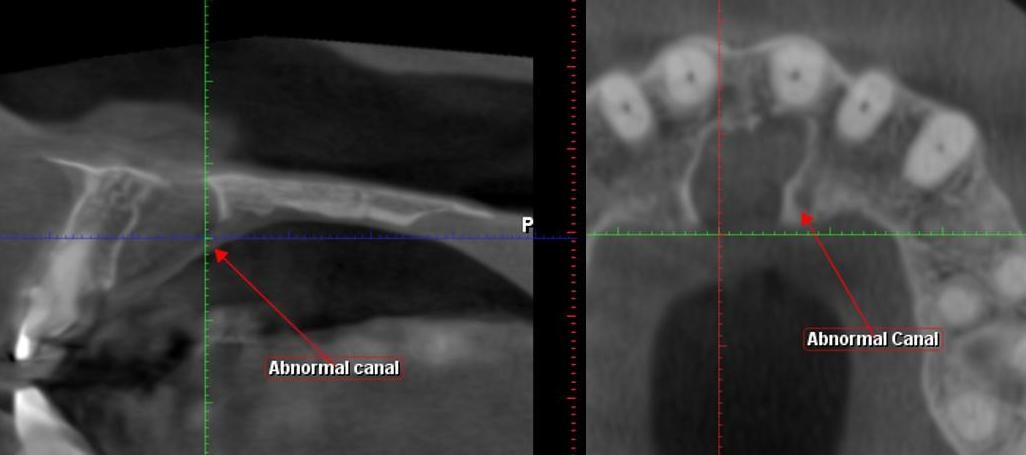
Example #2 of what a cyst looks like:
